
Page 33 - TPIS2022
P. 33
Lecture title:
Molecular Imaging with Positron Emission Tomography for Drug
Development in Pulmonary Fibrosis
ABSTRACT:
In recent years, micro-computed tomography (microCT) imaging has become increasingly
popular for assessing trabecular and cortical bone morphology in human and laboratory
animal specimens. Several studies have demonstrated that microCT measurements of bone
morphology are exceptionally reproducible and accurate, and are highly correlated with those
obtained from 2D histomorphometry. Use of microCT for bone analysis in excised specimens
has several advantages: It provides 3D measurements of trabecular morphology and thickness
at an enlarged volume of interest, faster than histologic analysis, nondestructive assessment,
and estimates mineralization within bone tissue. It has become the “gold standard” in
evaluating bone morphology and microarchitecture in mice and other small animals.
In this report, we describe three important applications of micro-CT scan imaging
in orthopedic translational research. First, Evaluation and standardization of a translational
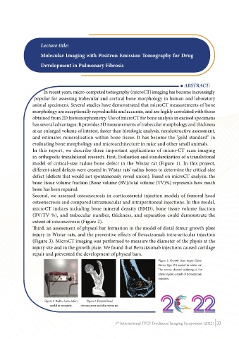
model of critical-size radius bone defect in the Wistar rat (Figure 1). In this project,
different-sized defects were created in Wistar rats’ radius bones to determine the critical-size
defect (defects that would not spontaneously reveal union). Based on microCT analysis, the
bone tissue volume fraction (Bone volume (BV)/total volume (TV)%) represents how much
bone has been repaired.
Second, we assessed osteonecrosis in corticosteroid injection models of femoral head
osteonecrosis and compared intramuscular and intraperitoneal injections. In this model,
microCT indices including bone mineral density (BMD), bone tissue volume fraction
(BV/TV %), and trabecular number, thickness, and separation could demonstrate the
extent of osteonecrosis (Figure 2).
Third, an assessment of physeal bar formation in the model of distal femur growth plate
injury in Wistar rats, and the preventive effects of Bevacizumab intra-articular injection
(Figure 3). MicroCT imaging was performed to measure the diameter of the physis at the
injury site and in the growth plate. We found that Bevacizumab injections caused cartilage
repair and prevented the development of physeal bars.
Figure 3. Growth plate injury (Salter
Harris type IV) model in wistar rat.
The arrows showed widening of the
physical plate a result of Bevacizumab
injection.
Figure 1. Radius bone defect Figure 2. Femoral head
model in wistar rat. osteonecrosis model in wistar rat.
5 International TPCF Preclinical Imaging Symposium (2022) 33
th